Pregnancy ultrasound examinations
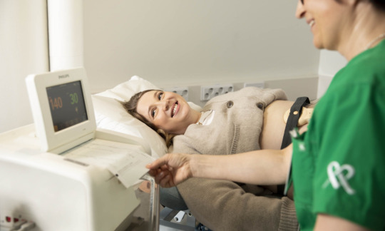
Early pregnancy ultrasound examination shows whether the foetus has settled in the correct place inside the uterus. It is also possible to determine the duration of the pregnancy and the due date of delivery in the examination.
Ultrasound examinations during pregnancy
Early pregnancy ultrasound examination
The early pregnancy ultrasound examination ensures that the foetus is in the right place inside the uterus. The examination reveals the number of foetuses and helps determine the duration of pregnancy.
An early pregnancy ultrasound examination can be performed during weeks 7–8 of pregnancy, when the foetus’s heart rate can already be observed.
Early pregnancy ultrasound examination is usually done through the vagina. Your bladder must be empty during the examination. An hour-based doctor’s fee is added to the fee of the ultrasound examination.
Nuchal fold ultrasound
The thickness of the foetus’s neck can be measured and the duration of pregnancy and the due time can be verified on the basis of the foetus’s crown-rump length during pregnancy weeks 11–13+6. The examination can be carried out either on the abdominal wall or through the vagina, depending on the pregnancy week in which it is performed.
Morphology scan
The purpose of a morphology scan carried out on pregnancy weeks 19–22 is to identify severe foetal structural abnormalities and to allow further examinations before week 24 of the pregnancy. The scan determines the number, condition, growth and morphology of foetuses, the amount of amniotic fluid and the location and structure of the placenta. A morphology scan carried out during these weeks of pregnancy leaves sufficient time for any possible further examinations, consultation and deciding on further measures.
After pregnancy week 24
After pregnancy week 24, ultrasound examinations are carried out to monitor the growth and condition of the foetus, placental function, amniotic fluid volume and cervical length.
3D/4D ultrasound examination
3D and 4D ultrasound examinations are three or four-dimensional ultrasound examinations. These examinations provide a multidimensional view of the foetus’s morphology, particularly the surface structures.
In a 3D/4D ultrasound examination, the foetus can be viewed both posteriorly and anteriorly, which allows the foetus’s facial features, spine and any related structural defects or tumours to be seen.
The foetus’s movements can also be seen in a three-dimensional view. Photos and videos can be saved in the OmaMehiläinen app for home viewing.
3D and 4D ultrasound examinations can be performed up to pregnancy week 30, if necessary. However, the best weeks for performing an ultrasound examination in terms of results and visibility are weeks 24–26.
3D/4D ultrasound examinations can also be performed in connection with other gynaecological examinations, as congenital abnormalities in the uterus, for example, can be detected more accurately than with normal ultrasound equipment.
Kela reimbursement for the examination must be applied for afterwards if it is carried out after pregnancy week 21.