3D/4D ultrasound examination
A 3D/4D ultrasound examination allows for a more multidimensional, accurate and reliable image of the foetus than with traditional ultrasound equipment.
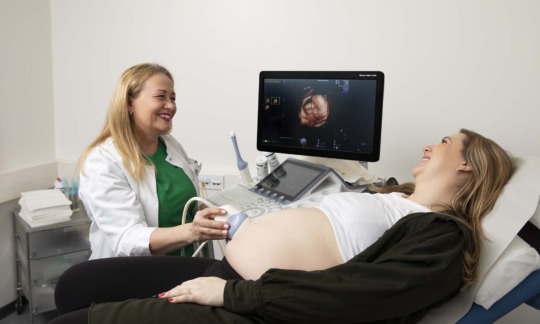
Ultrasound examinations with 3D/4D equipment
Felicitas Mehiläinen’s pregnancy monitoring specialists, the perinatologists, have access to a high-quality ultrasound device that produces three-dimensional video image of the foetus. This ultrasound equipment also provides a more accurate 2D image.
3D/4D ultrasound examinations allow some structural anomalies to be seen more accurately and reliably than in a traditional 2D ultrasound examination. In the examination, the foetus’s heart, brain, face, limbs, back and abdominal wall can be studied quite accurately.
In addition to its medical significance, three-dimensional ultrasound video imaging supports the relationship between the child and the parents already during pregnancy. In a 4D ultrasound examination, the baby’s features can be seen clearly if the foetus’s face is at an appropriate angle. An experienced doctor can also determine the gender of the child very reliably with the help of 4D ultrasound.
When should I book a 3D/4D ultrasound examination?
There is no specific limit by when you should have a 3D/4D ultrasound examination, but the view of the uterus may be narrow or limited during the later stages of pregnancy.
Obesity, the amount of amniotic fluid, an awkward position of the foetus and a placenta attached to the front wall can affect how well the uterus can be seen in an ultrasound examination.
In terms of visibility, the best pregnancy weeks for carrying out the examination are weeks 24–26, but the study can still be carried out around pregnancy weeks 28–30.
No separate referral is required for a 3D/4D ultrasound examination. A specific medical reason for the examination is required for Kela reimbursement. The Kela reimbursement is applied for afterwards. If anything abnormal is detected in the examination, the doctor who performed the ultrasound examination will guide you to the required further examinations on a case-by-case basis.
3D/4D ultrasound examinations can also be performed in connection with other gynaecological examinations, as congenital abnormalities in the uterus, for example, can be detected more accurately than with normal ultrasound equipment.
How does a 3D/4D ultrasound examination work?
A 3D ultrasound examination uses a three-dimensional image. 4D ultrasound, on the other hand, refers to a moving image, on the basis of which the 3D image is used. The two-dimensional image provided by Felicitas Mehiläinen’s 4D ultrasound device is also very accurate, as a 2D image of the highest quality is required for the imaging.
The 4D ultrasonic device calculates and models a three-dimensional image from small voxels. A voxel is a small unit of volume and it is not the same as a pixel, which many people are familiar with when talking about two-dimensional images.
In 3D/4D imaging, the sensor head motor of the ultrasound scanner moves the angle of view in three dimensions, which may feel like a small vibration on the stomach. The movement of the motor creates a three-dimensional image on the screen.
Photos and videos from the ultrasound examination can be saved for home viewing. The ultrasound examination session can also be recorded. The recording is available for a fee of a few euros.
3D/4D ultrasound examination price
The fee of a 4D ultrasound examination includes an appointment with a perinatologist, a gynaecologist specialised in pregnancy and childbirth, as well as the ultrasound examination. A pregnancy monitoring ultrasound examination can be performed at any stage of pregnancy if the pregnancy has progressed beyond pregnancy week 21.